


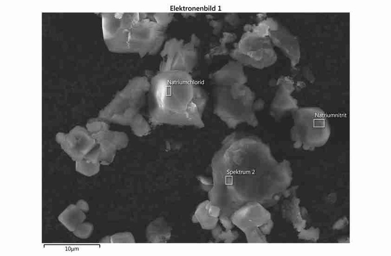
SEM - Scanning electron microscope
The surface of the specimen to be investigated is scanned with the aid of an electron beam. Secondary electrons (SE) are released from the surface of the specimen by the energy of the electron beam. These secondary electrons are used to illustrate the surface topography and make possible spatial resolution in the nanometre range.
Part of the primary electron beam is reflected back by the material of the specimen. The efficiency of the back-scattering effect thus depends on the density of the material - the denser the material, the more intensive the back-scatter. The detection of these back-scatter electrons (BSE) therefore provides a material contrast.
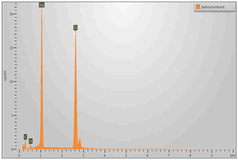
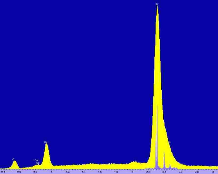
A large part of the energy of the electron beam leads to the excitation of the atoms of the material of the specimen, which leads to an emission of X-ray radiation. The characteristic proportion of X-ray radiation can be used for a chemical analysis of the specimen material (REM/EDX, REM/WDX). The determination of the chemical composition in accordance with ISO 22309 can therefore be done at a point, over an area, along a line (line scan) or scanning (element mapping).
SEM-Gallery


accreditation